Zellaufbau
Zeigt 13-24 von 43 Produkten 43 Produkte in Zellaufbau
Filters
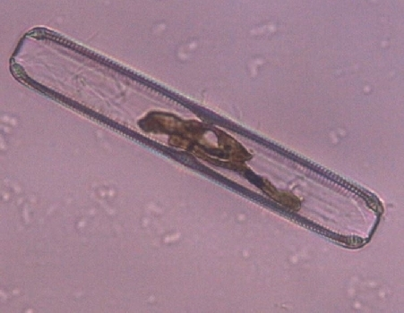
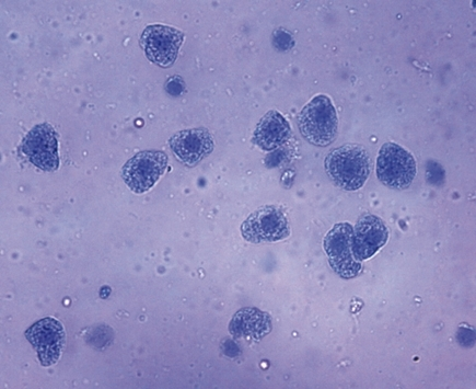
Kieselalgen im Moorwasser
Prinzip Viele Menschen fürchten sich vor dem gefährlichen Moor, weil man in ihm versinken kann. Für andere ist es aber ein überaus interessanter Ort. Archäologen finden im Moor Spuren von früher Besiedlung, Paläobotaniker suchen nach Pollen, die Pflanzen der Vorzeit hinterlassen haben. Botaniker finden hier besonders interessante fleischfressende Pflanzen und die Zoologen eine vielfältige Tierwelt. Das saure Wasser des Moores eröffnet uns auch mit dem Mikroskop eine andere Welt! Vorteile • Versuch ist Teil einer Komplettlösung mit insgesamt 50 Versuchen für alle Mikroskopanwendungen • Mit Schülerarbeitsblatt, das für alle Klassenstufen geeignet ist • Mit detaillierten Lehrerinformationen, inkl. Beispielmikroskopiebild • Besonders geignet bei knapper Zeitplanung, da minimale Vorbereitungszeit • Dazu passendes Mikroskopie-Set enthält alles für die Mikroskopie notwendige Zubehör • Dazu passende Multimediainhalte erhältlich mit unterrichtsbegleitenden Materialien
CHF 581.55
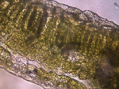
Laubblatt im Querschnitt
Prinzip Das Laubblatt einer Pflanze besteht aus verschiedenen Schichten. In den inneren Bereichen befinden sich Zellen mit vielen Chloroplasten. Die äußere Gewebeschicht, die Epidermis, schützt die Pflanze vor Verdunstung. Sie enthält keine Chloroplasten und die Zellen erscheinen unter dem Mikroskop durchsichtig. In der Palisadenschicht stehen dicht an dicht nach oben gestreckte Zellen, die sehr viele grüne Chloroplasten enthalten. Darunter folgt das Schwammparenchym und dann wieder die abschließende Epidermis. Vorteile • Versuch ist Teil einer Komplettlösung mit insgesamt 50 Versuchen für alle Mikroskopanwendungen • Mit Schülerarbeitsblatt, das für alle Klassenstufen geeignet ist • Mit detaillierten Lehrerinformationen, inkl. Beispielmikroskopiebild • Besonders geignet bei knapper Zeitplanung, da minimale Vorbereitungszeit • Dazu passendes Mikroskopie-Set enthält alles für die Mikroskopie notwendige Zubehör • Dazu passende Multimediainhalte erhältlich mit unterrichtsbegleitenden Materialien
CHF 844.30
Leberzellen
Prinzip Die Leber ist ein zentrales Stoffwechselorgan der Tiere und des Menschen. Sie beeinflusst z. B. den Blutzuckerspiegel, produziert verschiedene Bluteiweiße und baut giftige Stoffwechselprodukte und andere mit der Nahrung aufgenommene Gifte ab. Der von der Leber produzierte Gallensaft wird in der Gallenblase gesammelt und bei Bedarf in den Darm abgegeben. Er dient der Zerteilung des Nahrungsfettes. Die Leber des Menschen ist mit ca. 1500 Gramm ein sehr großes Organ und liegt im rechten Bauchbereich direkt unterhalb des Zwerchfells. Vorteile • Versuch ist Teil einer Komplettlösung mit insgesamt 50 Versuchen für alle Mikroskopanwendungen • Mit Schülerarbeitsblatt, das für alle Klassenstufen geeignet ist • Mit detaillierten Lehrerinformationen, inkl. Beispielmikroskopiebild • Besonders geignet bei knapper Zeitplanung, da minimale Vorbereitungszeit • Dazu passendes Mikroskopie-Set enthält alles für die Mikroskopie notwendige Zubehör • Dazu passende Multimediainhalte erhältlich mit unterrichtsbegleitenden Materialien
CHF 843.50
Meiosemodell
Vorteile der Modelle zur Meiose · Chromosomen nach modifizierter Azanfärbung gefärbt · Zellbestandteile nach didaktischen Gesichtspunkten gefärbt · Befestigungsmagnete an der Rückseite · Aufbewahrungssystem zum Stellen oder Hängen · Lieferung mit ausführlicher Beschreibung und Kopiervorlagen · 10.000-fache Vergrösserung Das dreidimensionale Reliefmodell zeigt die 10 Stadien der Meiose am Beispiel einer typischen Säugetierzelle: 1. Interphase (Stadium der G1-Phase) 2. Prophase I (Leptotän) 3. Prophase I (Zygotän und Pachytän) 4. Prophase I (Diplotän) 5. Prophase I (Diakinese) 6. Metaphase I 7. Anaphase I 8. Telophase I, Zytokinese I, Interkinese, Prophase II und Metaphase II 9. Anaphase II 10. Telophase II und Zytokinese II Abmessungen: ca. 60x40x6 cm³ Gewicht: ca. 1,7 kg
CHF 531.50
Mitose und Meiose I, Englisch
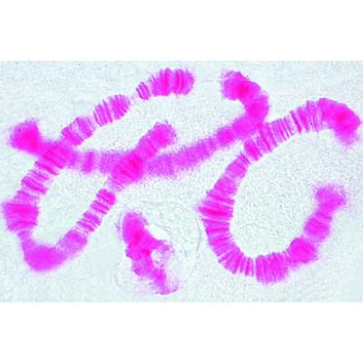
6 selected Microscope Slides. With depictured accompanying brochure 1(d). Mitosis, l.s. from Allium root tips showing plant mitosis stained with iron-hematoxyline 2(f). Mitotic stages in sec. of red bone marrow 3(e). Meiotic and mitotic stages in sec. of Salamandra testis 4(f). Lilium, anther t.s., microspore mother cells showing telophase of first and prophase of second division 5(f). Giant chromosomes, smear from salivary gland of Chironomus 6(f). Ascaris megalocephala embryology. Sec. of uteri showing maturation stages. 1013474 - Mitosis and Meiosis Set II
CHF 137.65
Mitose und Meiose I, Französisch
6 selected Microscope Slides. With depictured accompanying brochure 1(d). Mitosis, l.s. from Allium root tips showing plant mitosis stained with iron-hematoxyline 2(f). Mitotic stages in sec. of red bone marrow 3(e). Meiotic and mitotic stages in sec. of Salamandra testis 4(f). Lilium, anther t.s., microspore mother cells showing telophase of first and prophase of second division 5(f). Giant chromosomes, smear from salivary gland of Chironomus 6(f). Ascaris megalocephala embryology. Sec. of uteri showing maturation stages. French
CHF 137.65
Mitose und Meiose I, Portugiesisch
6 selected Microscope Slides. With depictured accompanying brochure 1(d). Mitosis, l.s. from Allium root tips showing plant mitosis stained with iron-hematoxyline 2(f). Mitotic stages in sec. of red bone marrow 3(e). Meiotic and mitotic stages in sec. of Salamandra testis 4(f). Lilium, anther t.s., microspore mother cells showing telophase of first and prophase of second division 5(f). Giant chromosomes, smear from salivary gland of Chironomus 6(f). Ascaris megalocephala embryology. Sec. of uteri showing maturation stages. Portuguese
CHF 137.65
Mitose und Meiose I, Spanisch
6 selected Microscope Slides. With depictured accompanying brochure 1(d). Mitosis, l.s. from Allium root tips showing plant mitosis stained with iron-hematoxyline 2(f). Mitotic stages in sec. of red bone marrow 3(e). Meiotic and mitotic stages in sec. of Salamandra testis 4(f). Lilium, anther t.s., microspore mother cells showing telophase of first and prophase of second division 5(f). Giant chromosomes, smear from salivary gland of Chironomus 6(f). Ascaris megalocephala embryology. Sec. of uteri showing maturation stages.SPANISH
CHF 137.65
Mitose und Meiose II, Deutsch
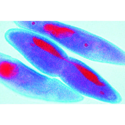
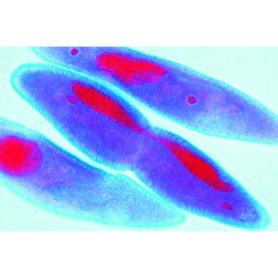
5 ausgewählte Präparate, mit ausführlichem Begleittext 1(d). Zellteilungen (Mitosen). in den Wurzelspitzen von Vicia faba, Bohne, längs 2(f). Lilium, Antheren quer. Pollenmutterzellen, Metaphase und Anaphase der ersten Reifungsteilung (Meiose) 3(h). Mitosestadien in der Keimscheibe eines Fisches mit Zentrosphären 4(f). Heuschrecke, Hoden, quer. Spermatogenese mit Meiose- und Mitose-Stadien 5(g). Pantoffeltierchen, Paramaecium, Teilungsstadien. 1013466 - Mitose und Meiose Serie I
CHF 130.40
Mitose und Meiose II, Englisch
5 selected Microscope Slides. With depictured accompanying brochure 1(d). Mitosis, l.s. from Vicia faba (bean). root tips showing all mitotic stages. Iron hematoxyline 2(f). Lilium, anther t.s., microspore mother cells showing telophase of first and prophase of second division 3(h). Mitotic stages in sec. of whitefish blastula showing spindles 4(f). Spermatogenesis with meiotic and mitotic stages, sec. of testis of grasshopper 5(g). Paramaecium, in fission, nuclei stained. 1013468 - Mitosis and Meiosis Set I
CHF 130.40
Mitose und Meiose II, Französisch
5 selected Microscope Slides. With depictured accompanying brochure 1(d). Mitosis, l.s. from Vicia faba (bean). root tips showing all mitotic stages. Iron hematoxyline 2(f). Lilium, anther t.s., microspore mother cells showing telophase of first and prophase of second division 3(h). Mitotic stages in sec. of whitefish blastula showing spindles 4(f). Spermatogenesis with meiotic and mitotic stages, sec. of testis of grasshopper 5(g). Paramaecium, in fission, nuclei stained. French
CHF 130.40
Mitose und Meiose II, Portugiesisch
5 selected Microscope Slides. With depictured accompanying brochure 1(d). Mitosis, l.s. from Vicia faba (bean). root tips showing all mitotic stages. Iron hematoxyline 2(f). Lilium, anther t.s., microspore mother cells showing telophase of first and prophase of second division 3(h). Mitotic stages in sec. of whitefish blastula showing spindles 4(f). Spermatogenesis with meiotic and mitotic stages, sec. of testis of grasshopper 5(g). Paramaecium, in fission, nuclei stained. Portuguese
CHF 130.40

 Versuche & Sets
