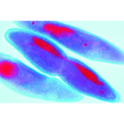
Ascaris megalocephata, Englisch

Artikelnummer: 1013479 [W13458]

CHF 171.-

inkl. MwSt. CHF 184.85

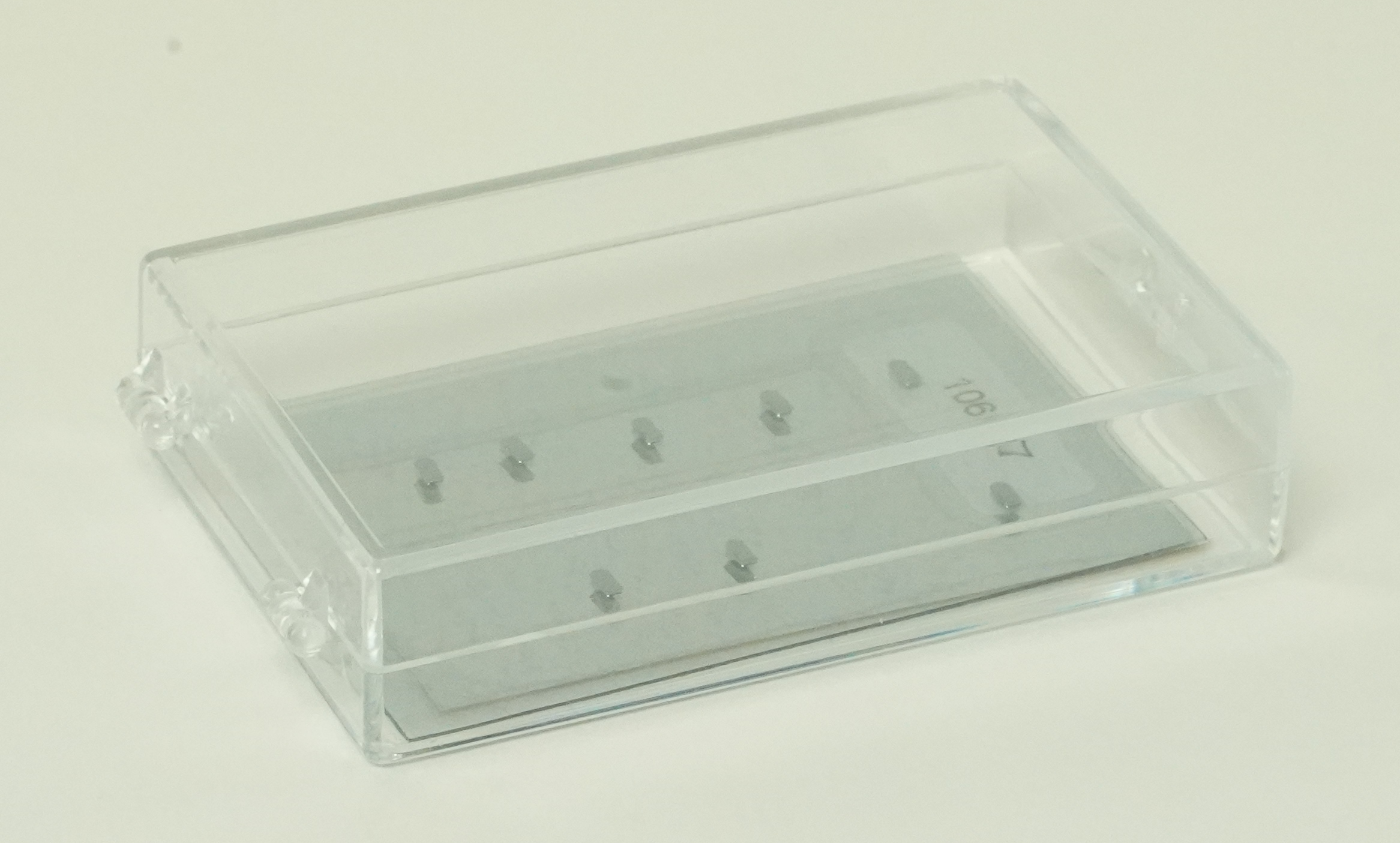
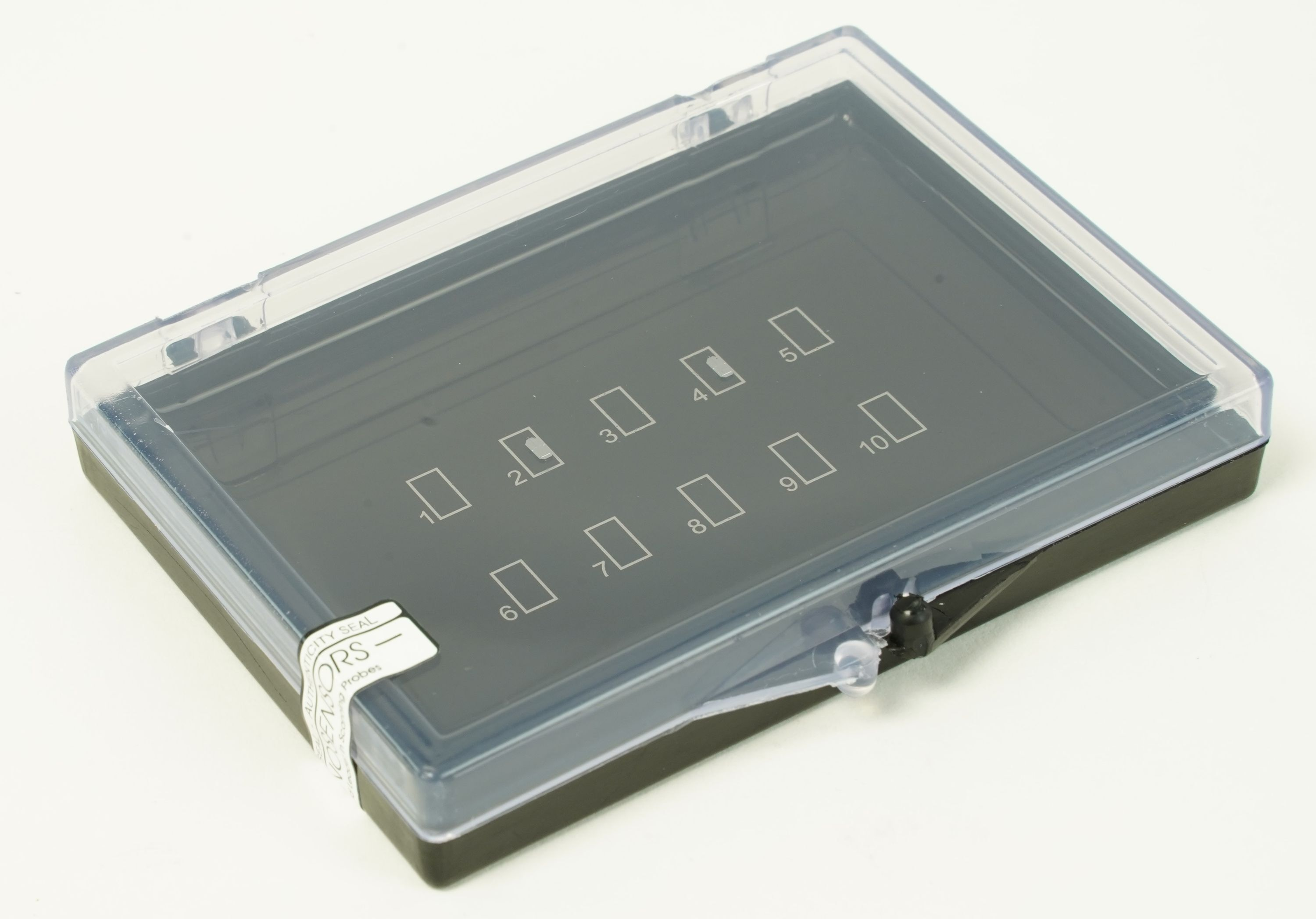
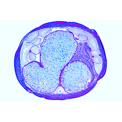
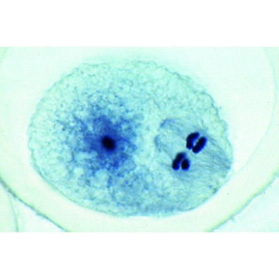
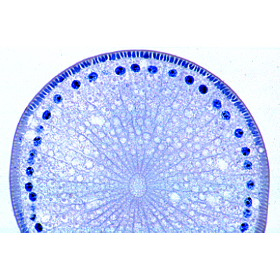
10 Microscope Slides. With depictured accompanying brochure 1(d). Cell division in l.s. of Allium root tips, showing all mitotic stages 2(e). Ascaris, primary germ cells in the growing zone of oviduct 3(f). Ascaris, entrance of sperm in the oocytes 4(f). Ascaris, first and second maturation divisions in oocytes I, 5(f). Ascaris, dito. in oocytes II 6(f). Ascaris, mature oocytes with male and female pronuclei 7(f). Ascaris, early cleavage stages 8(f). Ascaris, later cleavage stages 9(d). Ascaris, adult female, t.s. in region of gonads 10(d). Ascaris, adult male roundworm, t.s. in region of gonads.

10 Microscope Slides. With depictured accompanying brochure 1(d). Cell division in l.s. of Allium root tips, showing all mitotic stages 2(e). Ascaris, primary germ cells in the growing zone of oviduct 3(f). Ascaris, entrance of sperm in the oocytes 4(f). Ascaris, first and second maturation divisions in oocytes I, 5(f). Ascaris, dito. in oocytes II 6(f). Ascaris, mature oocytes with male and female pronuclei 7(f). Ascaris, early cleavage stages 8(f). Ascaris, later cleavage stages 9(d). Ascaris, adult female, t.s. in region of gonads 10(d). Ascaris, adult male roundworm, t.s. in region of gonads.
Ähnliche Produkte

  • CHF 241.20

  • Mitose und Meiose II, Französisch

    5 selected Microscope Slides. With depictured accompanying brochure 1(d). Mitosis, l.s. from Vicia faba (bean). root tips showing all mitotic stages. Iron hematoxyline 2(f). Lilium, anther t.s., microspore mother cells showing telophase of first and prophase of second division 3(h). Mitotic stages in sec. of whitefish blastula showing spindles 4(f). Spermatogenesis with meiotic and mitotic stages, sec. of testis of grasshopper 5(g). Paramaecium, in fission, nuclei stained. French

    CHF 130.40

  • Mitose und Meiose I, Portugiesisch

    6 selected Microscope Slides. With depictured accompanying brochure 1(d). Mitosis, l.s. from Allium root tips showing plant mitosis stained with iron-hematoxyline 2(f). Mitotic stages in sec. of red bone marrow 3(e). Meiotic and mitotic stages in sec. of Salamandra testis 4(f). Lilium, anther t.s., microspore mother cells showing telophase of first and prophase of second division 5(f). Giant chromosomes, smear from salivary gland of Chironomus 6(f). Ascaris megalocephala embryology. Sec. of uteri showing maturation stages. Portuguese

    CHF 137.65

  • Mitose und Meiose II, Deutsch

    5 ausgewählte Präparate, mit ausführlichem Begleittext 1(d). Zellteilungen (Mitosen). in den Wurzelspitzen von Vicia faba, Bohne, längs 2(f). Lilium, Antheren quer. Pollenmutterzellen, Metaphase und Anaphase der ersten Reifungsteilung (Meiose) 3(h). Mitosestadien in der Keimscheibe eines Fisches mit Zentrosphären 4(f). Heuschrecke, Hoden, quer. Spermatogenese mit Meiose- und Mitose-Stadien 5(g). Pantoffeltierchen, Paramaecium, Teilungsstadien. 1013466 - Mitose und Meiose Serie I

    CHF 130.40

  • Mitose und Meiose II, Englisch

    5 selected Microscope Slides. With depictured accompanying brochure 1(d). Mitosis, l.s. from Vicia faba (bean). root tips showing all mitotic stages. Iron hematoxyline 2(f). Lilium, anther t.s., microspore mother cells showing telophase of first and prophase of second division 3(h). Mitotic stages in sec. of whitefish blastula showing spindles 4(f). Spermatogenesis with meiotic and mitotic stages, sec. of testis of grasshopper 5(g). Paramaecium, in fission, nuclei stained. 1013468 - Mitosis and Meiosis Set I

    CHF 130.40

  • Mitose und Meiose II, Spanisch

    5 selected Microscope Slides. With depictured accompanying brochure 1(d). Mitosis, l.s. from Vicia faba (bean). root tips showing all mitotic stages. Iron hematoxyline 2(f). Lilium, anther t.s., microspore mother cells showing telophase of first and prophase of second division 3(h). Mitotic stages in sec. of whitefish blastula showing spindles 4(f). Spermatogenesis with meiotic and mitotic stages, sec. of testis of grasshopper 5(g). Paramaecium, in fission, nuclei stained. SPANISH

    CHF 130.40

  • Mitose und Meiose II, Portugiesisch

    5 selected Microscope Slides. With depictured accompanying brochure 1(d). Mitosis, l.s. from Vicia faba (bean). root tips showing all mitotic stages. Iron hematoxyline 2(f). Lilium, anther t.s., microspore mother cells showing telophase of first and prophase of second division 3(h). Mitotic stages in sec. of whitefish blastula showing spindles 4(f). Spermatogenesis with meiotic and mitotic stages, sec. of testis of grasshopper 5(g). Paramaecium, in fission, nuclei stained. Portuguese

    CHF 130.40

  • Meiosemodell

    Vorteile der Modelle zur Meiose ·         Chromosomen nach modifizierter Azanfärbung gefärbt ·         Zellbestandteile nach didaktischen Gesichtspunkten gefärbt ·         Befestigungsmagnete an der Rückseite ·         Aufbewahrungssystem zum Stellen oder Hängen ·         Lieferung mit ausführlicher Beschreibung und Kopiervorlagen ·         10.000-fache Vergrösserung Das dreidimensionale Reliefmodell zeigt die 10 Stadien der Meiose am Beispiel einer typischen Säugetierzelle: 1. Interphase (Stadium der G1-Phase) 2. Prophase I (Leptotän) 3. Prophase I (Zygotän und Pachytän) 4. Prophase I (Diplotän) 5. Prophase I (Diakinese) 6. Metaphase I 7. Anaphase I 8. Telophase I, Zytokinese I, Interkinese, Prophase II und Metaphase II 9. Anaphase II 10. Telophase II und Zytokinese II Abmessungen:        ca. 60x40x6 cm³ Gewicht:        ca. 1,7 kg

    CHF 531.50