Ergebnisse 13 – 20 von 20 werden angezeigt
Zellteilung
-
In den Warenkorb
Mitosis and Meiosis Set II
130.45 CHF
Mitosis and Meiosis Set II
5 selected Microscope Slides. With depictured accompanying brochure 1(d). Mitosis, l.s. from Vicia faba (bean). root tips showing all mitotic stages. Iron hematoxyline 2(f). Lilium, anther t.s., microspore mother cells showing telophase of first and prophase of second division 3(h). Mitotic stages in sec. of whitefish blastula showing spindles 4(f). Spermatogenesis with meiotic and mitotic stages, sec. of testis of grasshopper 5(g). Paramaecium, in fission, nuclei stained.
1013468 – Mitosis and Meiosis Set I
130.45 CHF -
In den Warenkorb
Mitosis and Meiosis Set II – French
130.45 CHF
Mitosis and Meiosis Set II – French
5 selected Microscope Slides. With depictured accompanying brochure 1(d). Mitosis, l.s. from Vicia faba (bean). root tips showing all mitotic stages. Iron hematoxyline 2(f). Lilium, anther t.s., microspore mother cells showing telophase of first and prophase of second division 3(h). Mitotic stages in sec. of whitefish blastula showing spindles 4(f). Spermatogenesis with meiotic and mitotic stages, sec. of testis of grasshopper 5(g). Paramaecium, in fission, nuclei stained.
French
130.45 CHF -
In den Warenkorb
Mitosis and Meiosis Set II – Spanish
130.45 CHF
Mitosis and Meiosis Set II – Spanish
5 selected Microscope Slides. With depictured accompanying brochure 1(d). Mitosis, l.s. from Vicia faba (bean). root tips showing all mitotic stages. Iron hematoxyline 2(f). Lilium, anther t.s., microspore mother cells showing telophase of first and prophase of second division 3(h). Mitotic stages in sec. of whitefish blastula showing spindles 4(f). Spermatogenesis with meiotic and mitotic stages, sec. of testis of grasshopper 5(g). Paramaecium, in fission, nuclei stained.
SPANISH
130.45 CHF -
In den Warenkorb
Mitosis and Meiosis Set II -Portuguese
130.45 CHF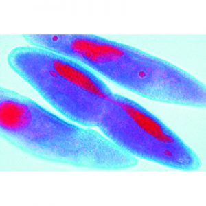
Mitosis and Meiosis Set II -Portuguese
5 selected Microscope Slides. With depictured accompanying brochure 1(d). Mitosis, l.s. from Vicia faba (bean). root tips showing all mitotic stages. Iron hematoxyline 2(f). Lilium, anther t.s., microspore mother cells showing telophase of first and prophase of second division 3(h). Mitotic stages in sec. of whitefish blastula showing spindles 4(f). Spermatogenesis with meiotic and mitotic stages, sec. of testis of grasshopper 5(g). Paramaecium, in fission, nuclei stained.
Portuguese
130.45 CHF -
In den Warenkorb
The Ascaris megalocephala Embryology
171.00 CHF
The Ascaris megalocephala Embryology
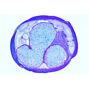
10 Microscope Slides. With depictured accompanying brochure 1(d). Cell division in l.s. of Allium root tips, showing all mitotic stages 2(e). Ascaris, primary germ cells in the growing zone of oviduct 3(f). Ascaris, entrance of sperm in the oocytes 4(f). Ascaris, first and second maturation divisions in oocytes I, 5(f). Ascaris, dito. in oocytes II 6(f). Ascaris, mature oocytes with male and female pronuclei 7(f). Ascaris, early cleavage stages 8(f). Ascaris, later cleavage stages 9(d). Ascaris, adult female, t.s. in region of gonads 10(d). Ascaris, adult male roundworm, t.s. in region of gonads.
171.00 CHF -
In den Warenkorb

The Ascaris megalocephala Embryology – French
10 Microscope Slides. With depictured accompanying
brochure
1(d). Cell division in l.s. of Allium root tips, showing all mitotic
stages 2(e). Ascaris, primary germ cells in the growing zone
of oviduct 3(f). Ascaris, entrance of sperm in the oocytes
4(f). Ascaris, first and second maturation divisions in oocytes
I, 5(f). Ascaris, dito. in oocytes II 6(f). Ascaris, mature oocytes
with male and female pronuclei 7(f). Ascaris, early cleavage
stages 8(f). Ascaris, later cleavage stages 9(d). Ascaris, adult
female, t.s. in region of gonads 10(d). Ascaris, adult male
roundworm, t.s. in region of gonads.171.00 CHF -
In den Warenkorb
The Ascaris megalocephala Embryology – Portuguese
10 Microscope Slides. With depictured accompanying
brochure
1(d). Cell division in l.s. of Allium root tips, showing all mitotic
stages 2(e). Ascaris, primary germ cells in the growing zone
of oviduct 3(f). Ascaris, entrance of sperm in the oocytes
4(f). Ascaris, first and second maturation divisions in oocytes
I, 5(f). Ascaris, dito. in oocytes II 6(f). Ascaris, mature oocytes
with male and female pronuclei 7(f). Ascaris, early cleavage
stages 8(f). Ascaris, later cleavage stages 9(d). Ascaris, adult
female, t.s. in region of gonads 10(d). Ascaris, adult male
roundworm, t.s. in region of gonads.171.00 CHF -
In den Warenkorb

The Ascaris megalocephala Embryology – Spanish
10 Microscope Slides. With depictured accompanying
brochure
1(d). Cell division in l.s. of Allium root tips, showing all mitotic
stages 2(e). Ascaris, primary germ cells in the growing zone
of oviduct 3(f). Ascaris, entrance of sperm in the oocytes
4(f). Ascaris, first and second maturation divisions in oocytes
I, 5(f). Ascaris, dito. in oocytes II 6(f). Ascaris, mature oocytes
with male and female pronuclei 7(f). Ascaris, early cleavage
stages 8(f). Ascaris, later cleavage stages 9(d). Ascaris, adult
female, t.s. in region of gonads 10(d). Ascaris, adult male
roundworm, t.s. in region of gonads.171.00 CHF